ECG vs 2D Echo vs Stress Echo vs Holter: Which Heart Test?
In brief
- An ECG records the heart's electrical signals in 5 to 10 minutes. It picks up rhythm problems, a fast or slow rate, and signs of strain or an old heart attack.
- A 2D echo is an ultrasound of the heart. It shows how strongly the heart pumps, how the valves work and whether the walls have thickened.
- A stress echo compares echo pictures at rest and after effort. It helps find narrowed heart arteries that a resting test can miss.
- A Holter monitor records every heartbeat for 24 to 48 hours at home, which suits palpitations or dizzy spells that come and go.
- Each test answers a different question, so your doctor may ask for one, two or none of them.
- Chest pain, sudden breathlessness or fainting is an emergency. Call 108 or 112.
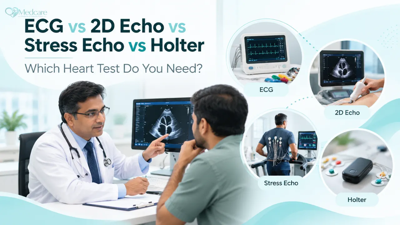
ECG vs 2D echo is one of the first things patients ask about once a heart test is mentioned. In short, the ECG reads the heart's electrical activity, while the 2D echo shows its structure and how well it pumps. A stress echo or Holter monitor comes in when those two still leave a question open. This guide covers what each test finds, what it misses, and what to expect on the day.
Four tests, four different questions
Patients often arrive with a printed ECG in one hand and an echo report in the other, wondering why one was "normal" and the other was not. The answer is that the two tests are looking at different things.
The heart has an electrical system that sets the beat, four chambers and four valves that move blood, a muscular wall that does the pumping, and its own arteries (the coronary arteries) that feed that muscle. An ECG reads the electrical system for a few seconds. A 2D echo looks at the chambers, valves and muscle while you rest. A stress echo watches the same muscle under effort, which tells us whether its blood supply keeps up. A Holter reads the electrical system again, only this time across a full day or two.
| Test | What it checks | Time taken | What you feel | Usually chosen for |
|---|---|---|---|---|
| ECG | Rhythm, rate, electrical signals | 5 to 10 minutes | Sticky pads on the skin, nothing more | Palpitations at the time, chest pain, a baseline in BP and diabetes |
| 2D echo | Pumping, valves, wall thickness, chamber size | 20 to 40 minutes | Cool gel, light pressure from the probe | Breathlessness, swollen feet, a murmur, years of high BP |
| Stress echo | How the muscle moves during effort | About 45 to 60 minutes overall | Tired legs, a fast heartbeat | Chest discomfort on walking or climbing, suspected blockage |
| Holter monitor | Rhythm over 24 to 48 hours or longer | Around 15 minutes to fit | A small recorder worn at home | Palpitations, dizziness or fainting that come and go |
What can an ECG tell you?
An ECG (electrocardiogram, sometimes written EKG) records the small electrical impulses that trigger each heartbeat. Ten pads go on the chest, wrists and ankles, and the machine prints a tracing within minutes. Nothing is passed into the body. The machine only records.
A careful look at that tracing can show atrial fibrillation (an irregular and often fast rhythm), a rate that is too quick or too slow, and delays in the heart's wiring known as heart block. It can also carry the marks of a recent or past heart attack, muscle thickening after years of high BP, or a potassium level that is too high or too low. For a test that costs little and takes minutes, it tells us a great deal.
What it cannot do is see beyond those 10 or so seconds. A patient whose heart races for half an hour every evening can have a perfectly normal ECG at 11 in the morning. And because narrowed arteries usually cause trouble only when the heart is working hard, the resting ECG in someone with significant blockage may look entirely normal.
When palpitations strike, note the time, what you were doing and how long they lasted. A few lines on your phone can save you a repeat test.
What is a 2D echo, and what does the report mean?
A 2D echo (two-dimensional echocardiogram) is an ultrasound of the heart, using the same kind of sound waves used in pregnancy scans. There is no radiation and, for a routine study, no injection. You lie on your left side, the doctor moves a probe over your chest, and the beating heart appears on the screen in real time.
Most people scan the report for one figure, the ejection fraction (EF). It is the share of blood the main pumping chamber, the left ventricle, pushes out with every beat. But the report covers much more. It notes whether the valves open fully and close without leaking, whether any chamber is enlarged, how thick the walls are, whether there is fluid in the sac around the heart, and an estimate of the pressure in the lung arteries.
There is also a line on diastolic function, which describes how well the heart relaxes and refills between beats. Readers tend to skip it, but in people with long-standing BP, diabetes or extra weight, this is often where the first change shows up.
Ejection fraction at a glance
| Ejection fraction | What it usually means |
|---|---|
| 50% or more | Pumping in the normal range, although the heart may still be stiff or thick |
| 41% to 49% | Mildly reduced pumping |
| 40% or less | Reduced pumping that needs regular specialist follow-up |
A normal EF does not close the matter. Someone can have an EF of 60%, swollen ankles and breathlessness on the stairs, all because the heart muscle has become stiff and does not fill well. Doctors call this heart failure with preserved ejection fraction. It is common in our part of the world among people living with diabetes and high BP, and it is easy to miss if only the EF is read.
Strain imaging
Strain imaging is an extra measurement taken during the echo. It tracks how much each part of the heart muscle shortens and lengthens as it beats. The muscle can begin to tire before the EF falls, and strain sometimes catches that early change in a person with diabetes or high BP while there is still time to protect the heart. Reference values differ from machine to machine, so the result is always read against the range for the equipment used.
One limitation should be clear. A resting echo does not show the coronary arteries, and a partly narrowed artery can leave the resting pictures looking normal.
When is a stress echo advised?
A stress echo answers the question a resting echo cannot, which is how the heart muscle behaves when it is asked to work harder. Pictures are taken at rest and again at peak effort. If an artery is narrowed, the wall it supplies may fall behind the rest of the heart during effort. That lagging segment, a wall motion abnormality, is what the doctor looks for.
Most people walk on a treadmill or pedal a stationary bicycle, with the effort increasing every few minutes. If knee pain, a weak back or age makes that difficult, a medicine such as dobutamine can be given through a drip to push the heart rate up instead. Either way, your BP, heart rate and ECG are watched from start to finish, and the test stops early if anything looks unsafe.
The classic reason to request one is chest tightness, heaviness or breathlessness that comes on with walking, climbing stairs or field work and fades with rest. It is also used when a resting ECG or echo raises doubt, to check some valve conditions under effort, and in selected patients before major surgery. A typical candidate might be a 55-year-old with diabetes, high BP and a smoking history who now gets short of breath walking uphill to the market.
People sometimes ask how this differs from the TMT (treadmill test) they had years ago. A TMT records only the ECG during exercise. A stress echo adds moving pictures of the heart, and for many patients that gives a far clearer answer.
It is not a test for an unstable moment. Chest pain at rest, a heart attack in the last few days, very high uncontrolled BP or a dangerous rhythm all need treatment first.
What does a Holter monitor catch?
A Holter monitor is a small recorder, about the size of a phone or smaller, wired to a few pads on your chest. You wear it home and go about your usual routine while it stores every single beat. Most recordings run for 24 to 48 hours, and longer monitoring is sometimes needed.
It earns its place when symptoms are occasional. It can pick up atrial fibrillation that lasts an hour and vanishes, extra beats that feel like a thud or a skip, brief pauses that leave you light-headed, and fast rhythms that switch on and off without warning. It also shows how your heart rate behaves through the day and during sleep.
The diary card that comes with it matters as much as the device. Each time you feel something, write the time and what you were doing. When the recording is analysed, your notes are matched against the trace. Feeling your heart race at 4 pm while the trace shows a normal rhythm at 4 pm is not a wasted test, because it tells the doctor the cause probably lies elsewhere.
Practically, bathe before the fitting since the recorder must stay dry, keep it on through the night, and live your normal day. A day spent lying in bed for fear of disturbing the wires gives a much less useful recording than an ordinary working day.
Matching your symptoms to the right test
There is no single best heart test. The right one depends on what the doctor needs to find out, and the plan may change after an examination.
| Your situation | Usual starting test | Possible next step |
|---|---|---|
| Palpitations happening now | ECG | Holter if the ECG is normal |
| Palpitations that come and go | ECG and Holter | Echo to check structure |
| Breathlessness or swollen feet | ECG and 2D echo | Blood tests and further tests based on findings |
| Chest tightness on walking or stairs | ECG and 2D echo | Stress echo or another test for blockage |
| High BP for several years | ECG | Echo to measure wall thickness |
| Diabetes for many years, no symptoms | ECG at regular review | Echo when there is a specific reason |
| Fainting or near-fainting | ECG | Holter, echo and others as needed |
| Murmur heard during a check-up | 2D echo | Repeat echo over the years if a valve issue is found |
You may notice that some tests are skipped. That is deliberate. A test done without a clear question tends to produce borderline findings, and borderline findings tend to produce more tests and more worry without changing treatment.
How diabetes, high BP and weight affect the heart
In clinics across Lower Assam, it is unusual to see one of these conditions on its own. Diabetes, high BP and extra weight tend to arrive together, and the strain they put on the heart adds up.
Diabetes speeds up narrowing of the arteries and raises the risk of heart attack and heart failure. Over the years it can also stiffen the heart muscle directly. A point many families do not realise is that a person with long-standing diabetes may not feel classic chest pain during a heart attack, because diabetes can dull the nerves that carry pain signals. The warning may instead be breathlessness, sweating, nausea, sudden tiredness or a vague discomfort high in the stomach that is easily put down to acidity.
That said, current diabetes guidance does not ask for a stress test in everyone with diabetes who feels well. The decision rests on symptoms, ECG changes and overall risk. Some guidance also suggests a blood test called BNP or NT-proBNP to look for early heart strain, followed by an echo if the level is raised.
High BP harms the heart more quietly. Pumping against higher pressure every day makes the heart wall thicken, much like an arm that lifts heavy loads for years. The thick wall slowly stiffens, and later it may weaken. An ECG is a standard first test for anyone withhigh blood pressure, and an echo measures that thickening more precisely.
Extra weight raises the heart's daily workload and is linked with atrial fibrillation. Untreated sleep apnea, in which breathing stops and restarts during sleep, adds more strain and pushes BP up, especially at night. Body size can make echo pictures harder to get and ECG signals smaller, and an experienced operator takes this into account.
Are heart tests safe?
ECG, echo and Holter recordings are among the safest tests in medicine, with no radiation and no needles. At most, the pads leave a little redness or itching that settles within a day or two.
A stress echo carries a small, real risk because the heart is pushed on purpose. During the test, chest discomfort, a drop or rise in BP, or an irregular rhythm can occur. This is exactly why it is done under supervision with monitoring throughout. People given dobutamine sometimes feel a pounding heartbeat, warmth in the face or a mild headache, which usually fade soon after the drip is stopped.
Preparing for your test
ECG. Skip body lotion or oil on the chest and limbs that morning so the pads stick properly. Sit quietly for a few minutes before the recording. A small patch of chest hair may need trimming.
2D echo. Eat normally and take your regular medicines unless told otherwise. Bring every old ECG and echo report you have. Comparing today's pictures with last year's often tells the doctor more than either report alone.
Stress echo. Have a light meal a few hours before, and wear walking shoes. Check with the doctor beforehand about your BP and heart medicines, as some of them blunt the heart rate response, and never stop a medicine on your own. If you have diabetes, ask how to time your meal and sugar medicines that day, and carry glucose tablets or a sweet in your bag.
Holter. Bathe beforehand, wear a loose top, and keep the diary card and a pen with you.
A front-opening shirt, kurta or blouse makes every one of these tests quicker and more comfortable.
Common misunderstandings about heart tests
A normal ECG means a healthy heart
It is good news, but it is a few seconds of recording taken at rest. Narrowed arteries, valve disease and intermittent rhythm problems can all hide behind a normal ECG.
An echo will show blockages
A routine echo does not show the coronary arteries at all. It can reveal muscle damaged by an old heart attack, but a blockage that causes trouble only on effort needs a stress test or a different investigation.
The Holter sends current into the heart
It does not. The recorder simply listens to the heart's own signals.
Heart tests are only needed for chest pain
Breathlessness, swollen ankles, palpitations, dizziness, and years of diabetes or high BP are all good reasons for a heart check. Many problems are picked up long before any pain.
A good report means the tablets can stop
More often it means the tablets are working. Stopping BP, sugar, cholesterol or heart medicines without advice can quickly undo that progress.
When should you see a doctor?
Make an appointment if breathlessness on walking is new or getting worse, if your ankles are swelling, or if palpitations, dizzy spells or chest heaviness on effort keep returning. Tiredness that feels out of proportion to your daily work deserves a mention too. If you have lived with diabetes or high BP for several years and never had a heart check, that is reason enough to ask.
Emergency signs: call 108 or 112
Do not wait for a booked test if you have:
- chest pain or pressure lasting more than a few minutes, especially with sweating, vomiting, or pain spreading to the arm, jaw, neck or back
- sudden severe breathlessness, or waking at night gasping for air
- fainting or collapse
- a very fast or irregular heartbeat with dizziness, chest pain or breathlessness
- sudden weakness of the face, arm or leg, or difficulty speaking
For anyone with diabetes, unexplained breathlessness, heavy sweating or upper stomach discomfort should be taken just as seriously.
At Medcare
Before suggesting any heart test, Dr. Das looks at your symptoms alongside your sugar, BP, weight and earlier reports, because the right test depends on the whole picture. ECG,echocardiography with strain imaging andHolter monitoring are done at the clinic in Bongaigaon, as is 24-hour ambulatory BP monitoring when office readings are not telling the full story. If a stress test or another investigation is needed, he will explain why and what comes next. Patients with a weak or stiff heart continue regular review throughheart failure care.
Key takeaways
- The ECG reads the heart's electrical activity and the 2D echo shows its structure, so one cannot stand in for the other.
- A stress echo is the usual next step when symptoms appear on effort and a blockage is suspected.
- A Holter monitor is the test for symptoms that come and go.
- People with diabetes, high BP or extra weight benefit from regular heart review, even without symptoms.
- Emergency symptoms need 108 or 112, not an appointment.
To go over your symptoms or reports,book an appointment with Dr. Das.
FAQs
Which is better, ECG or 2D echo?
Neither, because each looks at a different part of the heart. The ECG reads electrical activity in a few minutes and is easy to repeat. The echo shows the muscle, valves and pumping strength. Doctors usually begin with an ECG and add an echo when there is breathlessness, swelling, a murmur or high BP over many years. Plenty of patients need both.
Can a 2D echo detect heart blockage?
Not directly. A routine echo does not image the coronary arteries. It may show a section of muscle that moves poorly, often the mark of an old heart attack. A partly narrowed artery can cause trouble only during effort, and that is where a stress echo or another type of test becomes useful.
Do I need to fast before an echo?
For a resting echo, generally no. Eat breakfast and take your usual medicines unless the clinic says otherwise. A stress echo is different, as a heavy meal makes exercise uncomfortable, so keep it light. Ask in advance about heart and BP tablets, and if you have diabetes, about adjusting meal timing and sugar medicines on the day.
Can I take a bath while wearing a Holter monitor?
You should avoid it. Water can damage the recorder and loosen the pads, so have your bath just before the fitting. Wiping the lower body with a damp cloth is usually fine during the recording. If a pad comes off, call the clinic for advice instead of sticking it back on yourself.
I have diabetes but feel fine. Should I get my heart checked?
An ECG at regular review is common sense for most people with diabetes, especially after several years or when BP and cholesterol are also high. A stress test for everyone without symptoms is not recommended. Some patients may be offered a blood test for heart strain and an echo, depending on age, diabetes duration and other risks.
My ECG report says abnormal. How worried should I be?
The label on the printout comes from the machine, and it often flags minor or harmless variations. Some findings do need an echo or Holter to look further. A doctor will read the tracing alongside your symptoms and older ECGs before deciding. Keep the report safe for your visit, and seek urgent care only if you also have emergency symptoms.
Sources

Medically reviewed by Dr. Tamoghna Das, MBBS, MD (Medicine) · Assam Council of Medical Registration No. 20276 · Last reviewed 20 October 2026
Medical disclaimer. This article is for general information only. It is not a diagnosis and is not a substitute for a consultation with a doctor. For advice about your own health, please book an appointment. Read our medical disclaimer.
Seek urgent care now, call 108 or 112, or go to the nearest emergency department if you have chest pain, sudden breathlessness, fainting, weakness on one side of the body, very high blood sugar with vomiting or drowsiness, repeated low blood sugar with confusion, or a foot wound with redness, swelling or fever.